Function of the Diaphragm
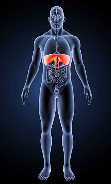
The diaphragm is the most important muscle involved in respiration . It is a dome-shaped sheet of tissue. It consists of both muscles and tendons. The diaphragm separates the chest cavity, containing the lungs and heart, from the abdominal cavity, containing organs such as the liver, kidneys, and gastrointestinal tract.
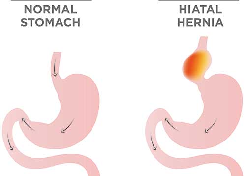
The diaphragm contains three major openings. The aortic hiatus is the opening through which the body’s largest artery (the aorta) passes from the heart into the abdominal cavity. Through the second opening, the inferior vena cava passes from the abdominal cavity into the thoracic cavity. The vena cava is firmly adherent to the diaphragm. The third opening allows the oesophagus to pass down to the stomach. This opening (termed the oesophageal hiatus) represents a natural weak point because the muscles in this region are comparatively lax. Particularly in cases where the diaphragm is weakened, parts of the stomach (or even the entire stomach!) can pass up through this gap and migrate into the chest cavity.In this case, the condition is referred to as a hiatal hernia, which is the pathological (abnormal) passage of parts of the stomach into the thoracic cavity.
Risk Factors
A hiatal hernia (i.e. diaphragmatic hernia) is the most common type of internal hernia. It can either be congenital (i.e. present from birth) or acquired later in life. In many cases, those affected also have a weakness of their connective tissues. When combined with further risk factors, the risk of a diaphragmatic hernia occurring increases.
Beyond age (most hiatal hernias occur after the age of 50), a particular risk factor is being severely overweight (i.e. obesity). Having previous abdominal surgeries can also predispose patients to developing a hiatal hernia.
Types of Diaphragmatic Hernia
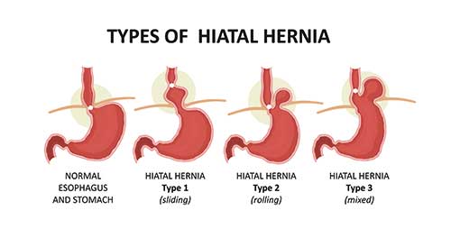
The most commonly seen hiatus hernia is termed an ‘axial-sliding’ hernia: this represents about 80-90% of all hiatus hernias.In this form, the junction between the oesophagus and stomach is displaced upwards into the thoracic cavity along the oesophagus.
In much rarer cases, a paraesophageal hernia may be seen (paraesophageal = adjacent to the oesophagus). In this type of hernia, parts of the stomach are displaced into the chest, while the junction between the oesophagus and stomach may remain in its original position or also be displaced into the chest, depending on the type of hernia. In more complex forms, other abdominal organs may also be displaced into the chest in addition to the stomach.
Key Symptom Heartburn
A hiatus hernia does not always cause symptoms. Many hernias, especially when small, remain asymptomatic long-term, and are often only discovered by chance. This can occur, for example, on X-ray imaging of the chest and/or abdomen. These hernias do not require treatment. We are faced with a different situation when the hernia causes symptoms, and as a result significantly impairs the patient’s quality of life.
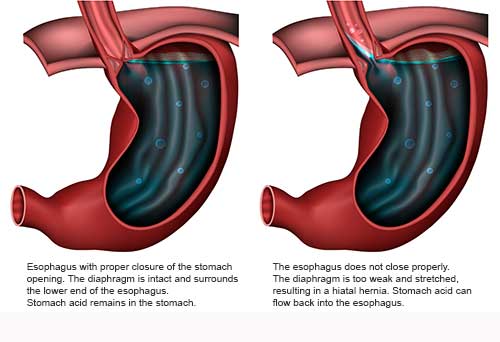
The most common clinical picture is termed gastroesophageal reflux disease (GERD), associated with severe and frequent heartburn, and often accompanied by difficulty swallowing and pain felt behind the breastbone – both symptoms tend to be worse when in a lying position. Furthermore, those affected may often suffer from a dry cough or asthma-type symptoms. Other possible symptoms which may occur include a burning sensation of the tongue, laryngitis, sleep apnoea, or even cardiac complaints – due to the heart’s ability to beat being impaired by the hiatal hernia. There has also been an increase in the worldwide incidence of oesophageal cancer – a possible consequence of a reflux disease which remains rare.
Reflux occurs when, due to a hiatal hernia, the lower oesophageal sphincter can no longer be supported by the diaphragm, allowing stomach acid to flow back into the oesophagus. In a healthy condition, this is prevented by the narrow opening in the diaphragm (oesophageal hiatus).